MSI hardware part 1: ionization techniques

Are you confused by all the acronyms used in mass spectrometry imaging? In this series of blogs, we hope to provide you with a brief introduction and explanation of some of the different things you might want to know about the technology, starting with the hardware. In this post, we will describe the main ionization techniques and tackle some of those pesky acronyms in the process.
This post is part of our series titled "Introduction to mass spectrometry imaging", which contains the following entries:
- MSI hardware part 1: ionization techniques (current post)
- MSI hardware part 2: mass analyzers
- MSI applications part 1: glycans
- MSI applications part 2: peptides and proteins
- MSI applications part 3: lipids
- MSI applications part 4: drugs and small molecule metabolites
Table of contents
Introduction
In the very first entry on this blog, we briefly explained that the anatomy of a mass spectrometer consists of three different parts: the ion source where analytes (molecules of interest) are ionized; the mass analyzer which separates the analytes based on their molecular weight; and the detector where the different analytes in the sample are detected. In this post we will introduce the most prevalent ionization techniques used in MSI.
To perform imaging, we need to somehow ionize analytes at specific locations in a tissue section, which rules out some ionization methods used in non-imaging MS. Nonetheless, a number of different ionization techniques are commonly used in MSI, which vary according to not only sample preparation requirements, but also measurement parameters, such as lateral resolution and mass range, and compatibility with other analytical techniques. Desorption ionization is most commonly used in MSI because it can be used to analyze a solid surface, and can be divided into three principal technologies: SIMS, MALDI and DESI. Schematics of each of these sources are shown in Figures 1 to 3 and a high-level comparison is provided in the comparison section below.
Secondary Ion Mass Spectrometry (SIMS)
Secondary ion mass spectrometry (SIMS) is the oldest of these technologies, being first used in the 1960s to measure elemental ions.1 In SIMS, the sample surface is sputtered with a primary ion beam which ablates material and causes the generation of secondary ions which are then analyzed.
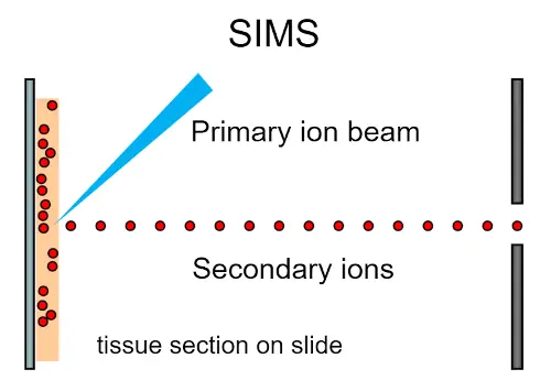
The source operates in an ultra-high vacuum so that primary ions can travel from the beam and secondary ions to the analyzer without colliding. An important sample preparation concern for SIMS imaging experiments is therefore stability during transport into the instrument for analysis under high vacuum. For tissue samples, this generally means mounting sections onto conductive glass slides. In addition, the surface of samples should be as flat as possible as rough surfaces can affect the generation of secondary ions and create shadow effects.
While a wide range of different samples and analytes can be analyzed by this technique, one of the major historical drawbacks of SIMS was the energy of the primary ion beam which can result in fragmentation of analyte molecules. As such, SIMS is generally not able to match other MSI technologies in the range of intact molecules detected. The surface of biological samples measured by SIMS can also be damaged which would then affect any further complementary analysis such as histological staining. Regardless, as ion beams can be focused with much higher precision than a laser beam, SIMS has the highest lateral resolution of current MSI ionization technologies for groups who are interested in subcellular molecular imaging.
An interesting feature of SIMS imaging is the ability to “dig into” tissue by repeatedly ablating material at the same location, providing a way to generate 3D MSI datasets from a single tissue section. Not surprisingly, such 3D datasets are often significantly larger than their 2D counterparts, which mandates appropriately scalable data analysis approaches.2
Matrix-assisted laser desorption/ionization (MALDI)
Matrix-assisted laser desorption/ionization (MALDI) is currently the MSI technique most commonly used for biological and biomedical applications. While direct irradiation of biological molecules with an intense laser pulse (LDI) can fragment the molecules of interest and lead to loss of structure, in MALDI biological molecules are mixed with a matrix – typically an organic acid – which absorbs the laser energy and aids molecular ionization.
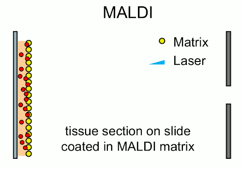
The use of MALDI for imaging tissue sections was first reported by Caprioli in 1997,3 who coined the term imaging mass spectrometry, essentially creating the field. MALDI as an MSI technology is extremely flexible as many different analytes can be detected with the method including but not limited to: proteins, peptides, glycans, lipids and drug compounds.
Compared to SIMS and DESI, sample preparation is extremely important in MALDI-MSI experiments and can be complex depending on the type of tissue sample and analyte of interest. For example, while measuring lipids from fresh frozen sections simply requires the sections be dried before matrix application, in order to measure peptides from formalin-fixed paraffin embedded (FFPE) tissue, the sections must undergo paraffin wax removal, rehydration, antigen retrieval and tryptic digestion.
Additionally, as most MALDI sources operate under either high or medium vacuum, sections must be mounted onto a conductive surface, usually a conductive glass slide. The use of glass slides means that MALDI is also compatible with other techniques such as histological staining for microscopic analysis or guiding further LC-MS/MS analysis.
Over the past decade, the speed at which a MALDI-based imaging experiment can be conducted has increased tremendously, with instruments now being capable of measuring full tissues in the order of minutes. This high throughput has made scalable data analysis increasingly important to handle the resulting large amounts of data.
Desorption electrospray ionization (DESI)
Desorption electrospray ionization (DESI) imaging is the newest of the ionization techniques discussed in this post, being reported in the mid-2000s. In DESI, the sample is sprayed with an electrically charged solvent mist (electrospray) which not only causes the ionization and desorption of analytes, but the direction of the electrospray to the sample surface also transports analytes into the mass spectrometer inlet.
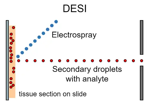
DESI, like ESI, generally generates ions with both single and multiple charges, whereas MALDI and SIMS usually results in only singly charged ions. Multiple charging is an important property with a characteristic impact on mass spectra that must be properly accounted for during data analysis.
Contrary to SIMS and MALDI, which operate under different levels of vacuum, DESI is performed at atmospheric pressure which allows for the analysis of solid, liquid and frozen samples, and generally requires little to no sample preparation, resulting in minimal damage to the samples, eliminates the potential of ionization interference from a matrix, and allows compatibility with other subsequent analyses. A negative aspect of operating at atmospheric conditions is that it has been reported that fluctuations in humidity can affect the stability of the electrospray.4 Moreover, DESI generally has poorer lateral resolution compared to SIMS and MALDI, however developments in hardware and data analysis techniques indicate that it can be improved upon.
Despite the relative youth of the technique, DESI has been demonstrated as useful for the characterization of lipid profiles of cancers and renal diseases, and for detecting small molecules, particularly drug compounds in-situ.
Comparison of ionization techniques
As explained in the previous sections, some fundamental differences exist between these ionization techniques which can have a large impact on their suitability for a given application, so comparing them head-to-head should be done with those differences in mind.
Nonetheless, it is also interesting to compare these techniques from a high-level perspective. Table 1 provides an overview of key properties for the ionization techniques described in this post.

The lateral resolutions we listed here are reported in 5 and 6 for MALDI, and 7 for DESI.
In this post, we introduced the main ionization techniques that are currently used for MSI. SIMS, MALDI and DESI MSI instruments are all commercially available from different vendors. While each of these ionization methods feature some unique properties, they all share the same goal of efficiently getting the analytes of interest ionized and funneled into a downstream mass analyzer.
Since the inherent structure of MS imaging data is largely equivalent across ionization methods, Aspect Analytics’ software can be used to analyze data that is generated using any of them. Our software is fully compatible with the open imzML format, which all vendors provide export tools for, as well as proprietary data formats of the leading vendors.
Stay tuned for the next installment in this series where we discuss the different mass analyzers available.
References
1. PortaSiegel, T. et al. “Mass Spectrometry Imaging and Integration with Other Imaging Modalities for Greater Molecular Understanding of Biological Tissues”. MolImaging Biol 20, 888-901, doi:10.1007/s11307-018-1267-y (2018).
2. Van Nuffel,S. et al. “Multivariate analysis of 3D ToF-SIMS images: method validation and application to cultured neuronal networks”. Analyst 141, 90-95, doi:10.1039/C5AN01743B (2015).
3. Caprioli,R. M., Farmer, T. B. & Gile, J. “Molecular imaging of biological samples: localization of peptides and proteins using MALDI-TOF MS”. Anal Chem 69,4751-4760, doi:10.1021/ac970888i (1997).
4. Feider, C.L. et al. “DESI Spray Stability in the Negative Ion Mode Is Dependent on Relative Humidity”. J Am Soc Mass Spectrom 30, 376-380, doi:10.1007/s13361-018-2105-9 (2019).
5. Zavalin,A., Yang, J., Hayden, K., Vestal, M. & Caprioli, R. M. “Tissue protein imaging at 1 mum laser spot diameter for high spatial resolution and high imaging speed using transmission geometry MALDI TOF MS”. Anal Bioanal Chem 407,2337-2342, doi:10.1007/s00216-015-8532-6 (2015).
6. Kompauer,M., Heiles, S. & Spengler, B. “Atmospheric pressure MALDI mass spectrometry imaging of tissues and cells at 1.4-mum lateral resolution”. Nat Methods 14,90-96, doi:10.1038/nmeth.4071 (2017).
7. Yin, R.,Burnum-Johnson, K. E., Sun, X., Dey, S. K. & Laskin, J. “High spatial resolution imaging of biological tissues using nanospray desorption electrospray ionization mass spectrometry”. Nat Protoc 14, 3445-3470, doi:10.1038/s41596-019-0237-4 (2019).